
SciBase Journals
SciBase Dentistry and Oral Sciences
ISSN 2996-363X
- Article Type: Case Report
- Volume 2, Issue 1
- Received: Mar 20, 2024
- Accepted: Apr 17, 2024
- Published Online: Apr 24, 2024
Mucocele in the Ventral Surface of the Tongue: A Case Report and Literature Review
Said Ahmad Sorosh Miri1*; Fariha Kamal2; Adamkhan Alipour3
1Department of Prosthodontic, Faculty of Stomatology, Khatam Al Nabieen University, Kabul, Afghanistan.
2Department of Oral Medicine, Stomatology Teaching Hospital, Kabul University of Medical Sciences, Kabul, Afghanistan.
3Department of Biochemistry, Faculty of Medical Laboratory Technology, Khatam Al Nabieen University, Kabul, Afghanistan.
*Corresponding Author: Said Ahmad Sorosh Miri
Department of Prosthodontic, Faculty of Stomatology, Khatam Al Nabieen University, Kabul, Afghanistan.
Email: sorosh.miri@knu.edu.af
Abstract
Citation: Miri SAS, Kamal F, Alipur A. Mucocele in the Ventral Surface of the Tongue: A Case Report and Literature Review. SciBase Dent Oral Sci. 2024; 2(1): 1011.
Introduction
Mucoceles form in the oral mucosa due to the buildup of saliva, resulting in the enlargement of the affected region [1]. These lesions display a distinct trait of regressing and reappearing in an alternating pattern. This can be attributed to the rupture of the cystic cavity and subsequent re-aggregation of saliva [2]. Following a rupture, these mucoceles can cause the emergence of painful ulcerations that typically heal within a matter of days [3]. Mucoceles are particularly prevalent in the oral mucosa, with a higher frequency of occurrence compared to other regions. However, due to the substantial number of cases that do not undergo histopathological examination, it is challenging to accurately determine the incidence of this lesion [4]. Mucoceles are categorized into two subcategories: mucus extravasation cysts, which are 90% prevalent and resemble pseudocysts due to lack of epithelial lining [2], and mucus retention cysts, which are true cysts formed due to obstruction of the salivary duct [3], with a 10% prevalence [2]. This study aimed to present our case and assess its clinical features and management in comparison to those described in the literature.
Case report
A 23-year-old female patient with a painless swelling under her tongue for one month, making eating and speaking difficult, was referred for diagnosis and treatment of a tongue lesion. The patient’s dental and medical histories were insignificant. Extraoral examination showed no cervical lymphadenopathy or neck asymmetry and revealed no swelling or lymphadenopathy. There had been no previous trauma. Upon intraoral examination, a lesion was discovered lateral to the midline on the ventral surface of the tongue and was a unilateral, mildly uncomfortable enlargement that was soft to the touch and blanched on digital pressure. The lesion was more than 20 mm in diameter, sessile-based, and bordered with thin mucosa (Figure 1). Although the lesion was asymptomatic, the patient reported difficulties with speech and mastication. The dental and medical histories of the patient were insignificant. The patient also mentioned shrinkage but could not recall any traumatizing incidents. The patient also mentioned shrinkage but could not recall any traumatizing incidents. A clear fluid was obtained through a needle aspiration, ruling out any lesions of vascular origin. The anatomical location of the lesion was suggestive of the involvement of the glands of Blandin and Nuhn, and the lesion was clinically identified as a mucocele.
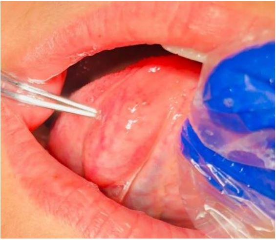
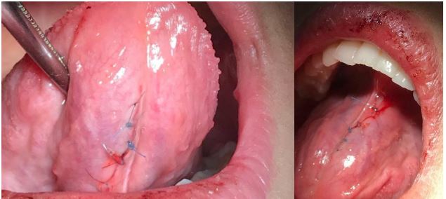
Table 1: Previously reported mucocele of the tongue.
| Author | Year | Patient’s age | Sex | Chief complaints | Location | Clinical finding |
|---|---|---|---|---|---|---|
| Basavaraj T Bhagawati, et al. [11] |
2021 | 7 years | Male | Swelling on the ventral surface of the tongue |
Ventral surface of the anterior tongue |
Fluctuant non-tender |
| Rayla Bentes Kato, et al. [12] |
2021 | 20 years | Male | Asymptomatic swelling | Midline of the ventral surface of the tongue |
Oval-shaped swelling with non-ulcerat- ed mucosa |
| Ashmitha J Rai, et al. [13] | 2007 | 8 years | Female | Unilateral painless swelling | Lateral of the ventral surface of the tongue |
Flaccid, sessile base with more than 15 mm |
| Saurabh R. Nagar, et al. [14] |
2020 | 11 years | Female | Painless swelling of the tongue | Ventral surface of tongue |
Painless, fluid-filled, soft, and solitary flaccid growth measuring about 10 mm × 8 mm |
| Thiago de Santana San- tos, et al. [15] |
2012 | 12 years | Male | Swelling on the ventral surface of the tongue |
Ventral surface of tongue |
A superficial protruding mass measuring 30×20 mm |
| Garcia Leon, et al. [16] | 2012 | 10 years | Female | Swelling on the ventral surface of the tongue with occasional pain |
Midline of the ventral surface of the tongue |
cystic, sessile, reddish mass of progres- sive growth |
| Praveen Kumar Pandey, et al. [17] |
2018 | 13 years | Female | Swelling on the lower surface of the tongue |
Ventral surface of tongue |
Bluish-red, non-tender fluid-filled mass measuring about 2 cm by 1cm |
| Jhon K Brooks, et al. [18] | 2016 | 22 years | Male | Painful mass on the ventral surface of the tongue |
Anterior midline ven- tral surface of tongue |
Firm, non-ulcerated pink-red umblicated papule |
| N. Graillon, et al. [19] | 2019 | Age range (7-39) |
4 Male/1 Female |
Swelling on the ventral surface of the tongue |
Ventral surface of tongue near frenulum |
Not included |
| Pandey PK, Gangwar S [17] |
2018 | 13 years | Female | Swelling on the lower surface of the tongue |
Ventral surface of the tongue |
Cystic fluid aspiration |
| Jose [20] | 2018 | 7 years | Female | Swelling in relation to the ven- tral surface of the tongue |
Ventral surface of the tongue |
pedunculated growth of size 10 mm × 5 mm |
| Titsinides [21] | 2018 | 74 years | Female | Painless swelling | Middle dorsal midline | Soft mass covered by mucosa 1.5 cm |
| Hur JH, et al. [22] | 2016 | 32 years | Female | No symptoms | Tongue midline | |
| 2021 | ||||||
| Our case | 2023 | 23 years | Female | Painless swelling on the ventral surface of the tongue |
Midline of the ventral surface of the tongue |
Soft, fluctuant, sessile-based swelling measuring more than 20 mm in size |
The final diagnosis of a mucocele cyst was made based on the clinical findings, and the patient was given the option of having the lesion surgically removed using standard surgical techniques. The lesion was excised under local anesthesia (Figure 2). The patient was given instructions regarding post-operative oral hygiene and care in the event of postoperative pain, analgesics were prescribed. The post-operative pain and edema were insignificant, and symptoms only lasted for a few days.
Discussion
Here, we presented a case of a mucocele in the ventral surface of the tongue in a 23-year-old female. Mucoceles are most commonly found on the lower lip, as it is prone to injury, followed by the floor of the mouth, the ventral tongue, and the buccal mucosa [1]. Mucoceles, which are found on the lower lip, are less common than on the anterior ventral surface of the tongue, where they are located near the lingual tonsils, Weber glands, and circumvallate papillae. They may also form on the base of clefts between foliate papillae and von Ebner serous salivary glands. Children are more likely to experience oral cavity pathologies, with mucous extravasation cysts accounting for 87.5% of cases [3]. To date, several cases of mucoceles have been reported with different Clinical ministrations. A collection of mucoceles of the tongue is shown in Table 1. Mucocele ranks as the 17th most prevalent salivary gland lesions observed within the oral cavity [5]. Clinically, mucocele are characterized by their spherical shape and fluctuant nature. They can appear as single or multiple lesions, ranging in color from a normal pink to a deep blue. Typically, they do not cause any symptoms. The deep blue color is a result of tissue cyanosis and vascular congestion, which is further accentuated by the stretched overlying tissue and the translucency of the accumulated fluid beneath. In some cases, these nodules may rupture, leading to slightly painful erosions. The most common location for extravasation mucoceles is the lower lip, which can be attributed to parafunctional habits such as lip biting and lip sucking, as well as trauma. These mucoceles are predominantly found in children and young patients, with equal incidences in both sexes. However, they are rarely seen in children under the age of one [6]. Although oral mucoceles rarely cause symptoms, they can occasionally interfere with speech, mastication, swallowing, and external swelling, as in our case [7]. The mucoceles that extend to the ventral surface of the tongue on the floor of the mouth are frequently misdiagnosed and confused with them [3]. Typically, mucoceles exhibit mobility and possess a soft and elastic consistency, which is contingent upon the amount of tissue covering the lesion. However, in the case of a drained mucocele, there would be no fluctuation, while a chronic mucocele accompanied by fibrosis would display reduced fluctuation. In retention-type mucoceles, a cystic cavity is observed, characterized by a distinct epithelial wall lined with cuboidal cells. This particular type demonstrates a lesser degree of inflammatory reaction. On the other hand, the extravasation type is akin to a pseudocyst, lacking an epithelial wall, and exhibiting inflammatory cells and granulation tissues. Despite the absence of an epithelial covering surrounding the mucosa, this type is well-encapsulated [8]. The diagnosis of a superficial mucocele is determined based on the historical and clinical evidence. The characteristic features of a mucocele, such as its location, history of trauma, sudden onset, fluctuating size, bluish hue, and texture, play a vital role in confirming the diagnosis [6]. The mucocele diagnosis in this study was based on the evaluation of factors including the anatomical site, size, flaccid nature, and asymptomatic nature of the lesion. Confirmation of the diagnosis and exclusion of salivary gland tumors necessitate the submission of the excised tissue for pathological investigations. In our specific case, the mucocele was excised and subsequently forwarded to the pathological department to validate the diagnosis. The conventional method for addressing this issue is to surgically remove the mucosa and glandular tissue below the muscle layer. While a simple incision of the mucocele can provide temporary relief by draining its contents, the lesion is prone to reappear once the wound has fully healed [5]. The main goal of treating mucocele is to completely remove the lesion to prevent recurrence. To prevent relapse, we must make sure that the diseased tissue, together with the affected and nearby glands, is eradicated [9,10].
Conclusion
Given that mucoceles are frequently disregarded or omitted, it is crucial to encompass mucoceles as a differential diagnosis for any observed growth on the midline of the ventral surface of the tongue in females during their second decade of life. Should this matter be neglected, it may result in further complications, such as excessive growth, that could hinder the process of mastication and interfere with swallowing. Superficial mucoceles may naturally regress and generally do not necessitate treatment; however, larger mucoceles must be surgically excised.
Declarations
Author contributions: Said ahmad sorosh miri: did the surgery, revised manuscript, led the study. Fariha Kamal: collected data. Adamkhan alipour: wrote the manuscript.
Consent: Written informed consent was obtained from the patient for publication of this case report and any associated images.
Funding statement: There has not been any financial support associated with this work.
Conflicts of interest: The authors declare that they have no conflict of unterest.
References
- More CB, Bhavsar K, Varma S, Tailor M. Oral mucocele: A clinical and histopathological study. Journal of oral and maxillofacial pathology: JOMFP. 2014; 18(Suppl 1): S72.
- Jani DR, Chawda J, Sundaragiri SK, Parmar G. Mucocele-A study of 36 cases. Indian Journal of Dental Research. 2010; 21(3): 337.
- Nagar SR, Fernandes G, Sinha A, Rajpari KN. Mucocele of the tongue: A case report and review of literature. J Oral Maxillofac Pathol. 2021; 25(Suppl 1): S37-s41.
- Plunkett L. A diagnostic dilemma. N Y State Dent J. 2010; 76(6): 6-7.
- Nallasivam KU, Sudha BR. Oral mucocele: Review of literature and a case report. J Pharm Bioallied Sci. 2015; 7(Suppl 2): S731-3.
- Bhargava N, Agarwal P, Sharma N, Agrawal M, Sidiq M, Narain P. An unusual presentation of oral mucocele in infant and its review. Case reports in dentistry. 2014; 2014.
- Andıran N, Sarıkayalar F, Ünal ÖF, Baydar DE, Özaydın E. Mucocele of the anterior lingual salivary glands: from extravasation to an alarming mass with a benign course. International journal of pediatric otorhinolaryngology. 2001; 61(2): 143-7.
- Rao PK, Hegde D, Shetty S, Chatra L, Shenai P. Oral mucocele–diagnosis and management. J Dent Med Med Sci. 2012; 2(2): 26-30.
- Navya L, Sabari C, Seema G. Excision of mucocele by using diode laser: a case report. Journal of Scientific Dentistry. 2020; 6(2): 30-5.
- Baburaj PAMD. Oral mucocoele: A case report. 2018.