
SciBase Journals
SciBase Epidemiology and Public Health
ISSN 2996-4555
- Article Type: Research Article
- Volume 2, Issue 1
- Received: Nov 15, 2023
- Accepted: Feb 06, 2024
- Published Online: Feb 13, 2024
Evaluation of Skin and Organ Dose of Patients Caused by Computed CT and Comparison with Monte Carlo Simulation Software GEANT4 (GATE)
Ghazizadeh E1,2; Ali Neshastehrizb2*
1Department of Bioinspired Materials and Biosensor Technologies, Institute of Materials Science, Faculty of Engineering, Kiel University, Germany.
2Department of Radiation Sciences, Iran University of Medical Sciences (IUMS), Tehran, Iran.
*Corresponding Author: Ali Neshastehrizb
Department of Radiation Sciences, Iran University of Medical Sciences (IUMS), Tehran, Iran.
Email: neshastehriz.a@iums.ac.ir
Abstract
Today, the use of CT scan as a type of diagnostic tool has increased dramatically. Therefore, controlled use and in accordance with protective regulations in order to reduce the harmful effects of radiation, it is necessary. The purpose of this study was to measure the dose received by patients in computed CT scan protocols and compare it with Monte Carlo simulation using GEANT4 software. Radiation parameters were collected from 11 patients referred to Tohid Hospital in Sanandaj to measure DLP quantity in common protocols. In this study, DLP values for Chest Abdomen protocol were measured and compared with simulation values. Our results show Monte Carlo software outputs experimental data well and is a good benchmark for this software. Thus, the simulated and measured doses agreed well.
Keywords: Computed Tomography; Chest CT scan; Monte carlo; Dose during scan; Reference dose limit.
Citation: Ghazizadeh E, Neshastehrizb A. Evaluation of Skin and Organ Dose of Patients Caused by Computed CT and Comparison with Monte Carlo Simulation Software GEANT4 (GATE). SciBase Epidemiol Public Health. 2024; 2(1): 1016.
Introduction
CT scan is an advanced imaging technique that provides cross-sectional and transverse images of body parts using X-rays using computer algorithms and calculations [1]. Today, use of CT scan as a type of diagnostic tool has increased dramatically. Specific information is required including activity distribution and organ boundaries for patient-specific dosimetry. CT data provides anatomical information which can be used for defining volume of interests specifying internal organs [2,3]. Nevertheless, using CT images for segmentation of anatomic structures of patient body, despite being more accurate, is time consuming. The alternative is using phantoms or Atlas data with already segmented organs and known organ boundaries. The anatomical structures are derived from these databases very easily [4].
In the United Kingdom, CT scans ranged from 250,000 to about 5 million from 1980 to 2013, representing a 20-fold increase, while in the United States, CT scans ranged from 2 million to 85 million. It has been shown to show a growth of approx. 43 [5]. In the United Kingdom and the United States, CT scans account for 11% and 17% of all medical X-ray tests and 67% and 49% of the cumulative effective dose, respectively. Absorption dose in tissues in CT scan is a higher component of the doses received by patients in diagnostic radiology methods [6,7]. Different parameters affect the dose received by patients in CT imaging. One of the most important factors influencing the dose received by patients is the intensity of the current in the tube (current generated in the tube due to the flow of electrons inside it) as a determinant of the amount of X-rays. For dosimetry calculations GATE (GEANT4) application to Tomographic Emission) [8], a Monte Carlo based script interface dedicated to nuclear medicine, was used. Different versions of this free open source toolkit are available on the open GATE collaboration website [9]. For dosimetry applications, GATE is capable to take either patient’s CT or a digital atlas phantom as input [10]. GATE has certain attractive features; some of them are inherited from GEANT4 [11] and some are additionally developed. These include flexible simulation geometry capable of accommodating a large variety of detector and source details and the physical events. In this study we review the evaluation skin and organ dose of patients caused by CT scan and comparison with Monte Carlo simulation software GEANT4 using DLP index.
Methods
Patient study: This study was performed on 11 patients referred to Tohid Hospital in Sanandaj for chest CT scan.
GE Light Speed RT, a third generation standard radiotherapy CT (GE Medical Systems, Milwaukee WI), was used in this study. The scanner has a large bore (80 cm), distance X-ray tube and isocenter 60.6 cm and performs 4-slice helical scanning. The tube voltage 80-140 kV step 20, tube current 10-440 mA step 5, rotation times of 1, 2, 3 and 4 seconds are available. Images were acquired with slice thicknesses of 2.5 mm on 10.0 mm collimation (4×2.5 mm) (GE Light Speed RT CT scanner technical evaluation November 2005). This scanner is used routinely for obtaining patient images for radiotherapy treatment planning at the Akdeniz University School of Medicine Department of Radiation Oncology. The regular quality assurance (QA) for image quality, 120-200 kV-mA measurement and mechanical tests based on national and international processes was performed. Three different body regions of the Rando phantom (head, chest and pelvic) were scanned by applying typical clinical protocols. The scan parameters kV, mA, pitch, FOV (field of view), rotation time, slice thickness of the CT examinations which were used in this study are given in Table 1. The scan length for each scanning protocol is also shown in Figure 1.
Monte carlo simulation: For both simulations of patientspecific dosimetry with the CT and XCAT phantom, the simulations were performed in GATE Monte Carlo code (version 6.0.0). The data of SPECT, CT and XCAT phantoms were processed to prepare suitable input file formats for GATE. The results of the internal dosimetry for the real activity distribution in the patient body based on the computed CT data were calculated for the CT image and the XCAT phantom in skin as well as in the total body. Photon absorption, Compton and Rayleigh scattering, ionizations, multiple scattering photons were simulated. After completion of simulations, GATE produced two binary files, containing respectively the absolute absorbed dose delivered into the voxels as DLP index (mGy) and the corresponding uncertainties [12]. Dycom photos of each case with VV the 4D slicer software converted into an MHA file or in another way with Mimics Medical 21.0 software converted to 3D STL files. Then, dosimetry separate programs were written for each of these inputs, in MHA and STL formats, and the output of both was almost the same, but in the 2D mode the results were closer to reality.
Dosimetry calculations: Dose Length Product (DLP) measured in mGy*cm is a measure of CT tube radiation output/ exposure. It is related to volume CT dose index (CTDIvol), but CTDIvol represents the dose through a slice of an appropriate phantom. DLP accounts for the length of radiation output along the z-axis (the long axis of the patient).
DLP = (CTDIvol)* (length of scan, cm) [units: mGy*cm]. DLP does not take the size of the patient into account and is not a measure of absorbed dose. If the AP and lateral dimensions of the patient are available, then the size specific dose estimate (SSDE) can be used to estimate the absorbed dose.
It is important to remember that the dose length product is not the patient’s effective dose. The effective dose depends on other factors including patient size and the region of the body being scanned. Some multipliers, called k-factors, have been estimated to convert DLPs into effective doses, depending on the body region. If interested, consult reference.
Table 1: Quality control tests include the accuracy and reproducibility of the parameters of each scan.
| L (cm) | I (mm) | T (mm) | P | mAs | KVp | Mode | protocol |
|---|---|---|---|---|---|---|---|
| 33.26-1.5 | 10 | 10 | 1.5 | 200 | 120 | helical | Breast |
Results
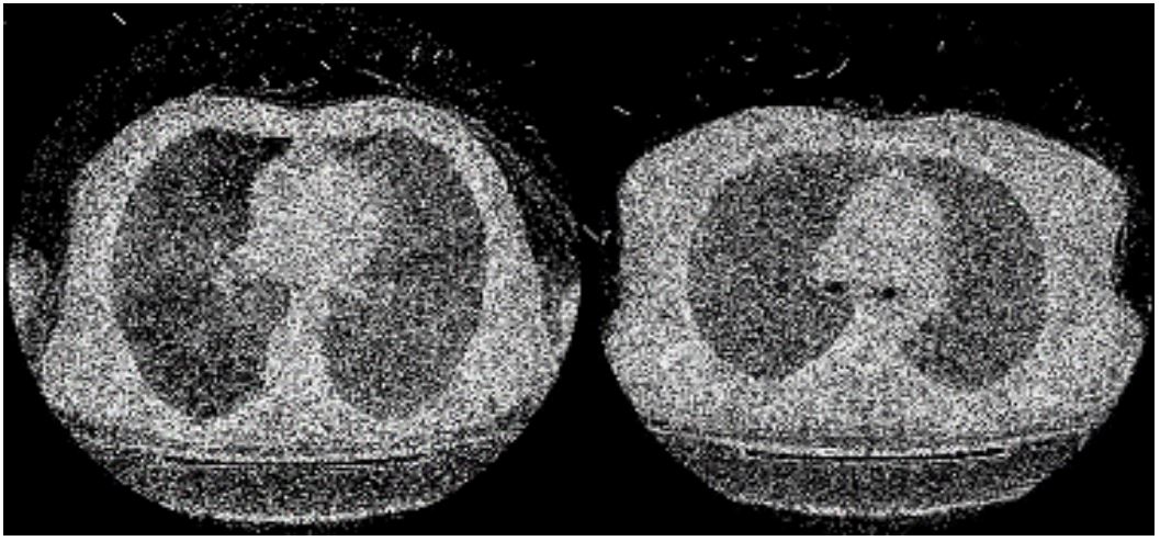
Organ dose simulations were performed using the scan parameters for the chest and abdomen–pelvis CT examinations. The scan range used for the chest CT contained the entire pulmonary area and that used for the abdominal–pelvic CT extended from the diaphragm to the pubic symphysis. In each simulation. The obtained results of DLP values for the dedicated GE Light Speed RT CT scanner for organ were about 250 mGy (Table 2). The reported values by manufacturer are 30.16 mGy and 23.9 mGy (GE Report 2005) so Commutated CT is used these values as standards at spreadsheet. The obtained results of DLP values from this study were less then reported values. As a general in the literature, the DLP value for conventional CT scanner is reported to be from 17 to 48 mGy [14,15]. For this dedicated CT scanner, the DLP values were in the range of values from conventional CT. In this study, the organ dose values were obtained by another measurement using the GATE Monte Carlo code (version 6.0.0) calculator and the two methods were compared for each scan protocol. The organs that were in the scanned region are blind listed in Table 3. First result of this study showed that the organ dose is relatively higher in helical mode by using GATE Monte Carlo simulation scanning.
Table 2: Results about dosimetry based on computed CT.
| Mode | DLP (mGy-cm) | Number |
|---|---|---|
| Helical | 259.9 | 1. W |
| Helical | 226.5 | 2. M |
| Helical | 248.7 | 3. -W |
| Helical | 231.6 | 4. -M |
| Helical | 247.3 | 5. -M |
| Helical | 258.3 | 6. e-M |
| Helical | 241.6 | 7. -M |
| Helical | 230.5 | 8. -M |
| Helical | 259.7 | 9. -W |
| Helical | 255.9 | 10. -M |
| Helical | 244/3 | 11. -M |
| Helical | 243/9 | 12. -W |
Table 3: Comparison between dosimetry based on CT and GATE Monte Carlo simulation.
| (DLP) GATE Monte Carlo code | DLP (CT Scan) | Number |
|---|---|---|
| 267.4 | 259.9 | 1 |
| 232.5 | 226.5 | 2 |
| 296.1 | 248.7 | 3 |
| 264.7 | 231.6 | 4 |
| 270.8 | 247.3 | 5 |
| 280.5 | 258.3 | 6 |
| 255.2 | 241.6 | 7 |
| 266.3 | 230.5 | 8 |
| 298.5 | 259.7 | 9 |
| 276.5 | 255.9 | 10 |
| 264.9 | 244.3 | 11 |
| 282.3 | 243.9 | 12 |
Discussion
We observed similar organ dosimetry results based on phantom with and patient’s CT data (Table 2). The similarity of the whole body dosimetry shows that the phantom and the calculation/simulations are generally acceptable. Variation between the organ boundaries and geometry of organs between patient and phantom may cause the differences and affect the organ dosimetry. In this study we used the GATE Monte Carlo code for calculation of absorbed dose. GATE code is already validated for dosimetry in many clinical situations including brachytherapy, external beam radiotherapy with photons/electrons, systemic radiotherapy, and proton-therapy. One of the main privileges of GATE is the capability to support both imaging and therapy modeling procedures [16]. The method we used has been employed with variations in other studies [17] for example to study mathematical phantom derived from the MIRD-type adult phantom. The use of phantoms is already validated for internal dosimetry purposes [15,12]. Another reports showed that the dosimetry based on phantom is different from those based on the Zubal phantom [27] as well as different dosimetry estimations obtained from different BMIs [28,29]. We showed, the calculated doses have a good approximation in the simulated software and the higher percentage of dose in the simulation can be attributed to the use of this approximation that the use of mono energy source in the simulated CT scan. The energy spectrum of the tube is not mono, and in a wide spectrum with a peak of one-third of energy, it sleeps like a rabbit. So in general, the computational results of DLS were similar.
Conclusion
In this study, we showed that the results of dosimetry similar when the CT phantom is used in place of patient’s CT image and GATE Monte Carlo code simulation. Providing a simulation method could be an option to give less right to CT scams.
References
- Grimes J, Celler A, Birkenfeld B, Shcherbinin S, Listewnik MH, Piwowarska-Bilska H, Mikolajczak R, Zorga P. Patient-specific radiation dosimetry of 99mTc-HYNIC- Tyr3-octreotide in neuroendocrine tumors. J Nucl Med. 2011; 52(9): 1474-81.
- Kolbert KS, Sgouros G, Scott AM, Bronstein JE, Malane RA, Zhang J, Kalaigian H, McNamara S, Schwartz L, Larson SM. Implementation and evaluation of patient- specific three-dimensional internal dosimetry. J Nucl Med. 1997; 38(2): 301-8.
- Saeedzadeh E, Sarkar S, Abbaspour Tehrani-Fard A, Ay MR, Khosravi HR, Loudos G. 3D calculation of absorbed dose for 131I-targeted radiotherapy: A Monte Carlo study. Radiat Prot Dosimetry. 2012; 150(3): 298-305.
- Sgouros G, Kolbert KS, Sheikh A, Pentlow KS, Mun EF, Barth A, Robbins RJ, Larson SM. Patient-specific dosimetry for 131I thyroid cancer therapy using 124I PET and 3-dimensional-internal dosimetry (3D-ID) software. J Nucl Med. 2004; 45(8): 1366-72.
- Tsougos I, Loudos G, Georgoulias P, Theodorou K, Kappas C. Patient-specific internal radionuclide dosimetry. Nucl Med Commun. 2010; 31(2): 97-106.
- Dewaraja YK, Frey EC, Sgouros G, Brill AB, Roberson P, Zanzonico PB, Ljungberg M. MIRD pamphlet No. 23: quantitative SPECT for patient-specific 3-dimensional dosimetry in internal radionuclide therapy. J Nucl Med. 2012; 53(8): 1310-25.
- Buck AK, Nekolla S, Ziegler S, Beer A, Krause BJ, Herrmann K, Scheidhauer K, Wester HJ, Rummeny EJ, Schwaiger M, Drzezga A. SPECT/CT. J Nucl Med. 2008; 49(8): 1305-19.
- Segars WP, Sturgeon G, Mendonca S, Grimes J, Tsuen BMW. 4D XCAT phantom for multimodality imaging research. Med Phys. 2010; 37(9): 4902-15.
- Bauman G, Charette M, Reid R, Sathya J. Radiopharmaceuticals for the palliation of painful bone metastasis-a systemic review. Radiother Oncol. 2005; 75(3): 258-70.
- Taschereau R, Chow PL, Cho JS, Chatziioannou AF. A microCT Xray head model for spectra generation with Monte Carlo simulations. Nucl Instrum Methods Phys Res A. 2006; 569(2): 373-377.
- Parach AA, Rajabi H. A comparison between GATE4 results and MCNP4B published data for internal radiation dosimetry. Nuklearmedizin. 2011; 50(3): 122-133.
- Díaz-Londoño G, García-Pareja S, Salvat F, Lallena AM. Monte Carlo calculation of specific absorbed fractions: variance reduction techniques. Phys Med Biol. 2015 7; 60(7): 2625-44.
- Fallahpoor M, Abbasi M, Kalantari F, Parach AA, Sen A. Practical Nuclear Medicine and Utility of Phantoms for Internal Dosimetry: XCAT Compared with Zubal. Radiat Prot Dosimetry. 2017; 174(2): 191-197.
- Fallahpoor M, Abbasi M, Parach AA, Kalantari F. Internal dosimetry for radioembolization therapy with Yttrium-90 microspheres. J Appl Clin Med Phys. 2017; 18(2): 176-180.
- Fallahpoor M, Abbasi M, Parach AA, Kalantari F. The importance of BMI in dosimetry of 153Sm-EDTMP bone pain palliation therapy: A Monte Carlo study. Appl Radiat Isot. 2017; 124: 1-6.
- Parach AA, Rajabi H, Askari MA. Paired organs-should they be treated jointly or separately in internal dosimetry? Med Phys. 2011; 38(10): 5509-21.
- Loevinger R, Budinger TF, Watson EE. MIRD primer for absorbed dose calculations. New York: Society of Nuclear Medicine. 1988.